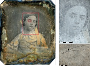
IN DETAILED studies for a heritage conservation project, researchers at the University of Antwerp in Belgium have found that high-resolution optical microscopy can be as powerful as scanning electron microscopy (SEM).
This is the claim made by Olympus, whose DSX500 Opto-digital system has been employed by a research team led by Dr Olivier Schalm at the Department of Conservation Studies at Antwerp.
It says that the cutting-edge digital imaging technologies and retention of true colour information at magnifications of up to 4000x has made this approach at least as fast and efficient as SEM.
Light microscopy is a popular analytical tool in cultural heritage researchs, gathering valuable information on the degradation of historic artefacts such as antique glass and daguerreotype photographic plates.
Only by truly understanding the mechanisms of degradation can one develop and optimise techniques to control and slow the process, or restore an object to a past state.
Such aims form the major focus of Dr Schalm’s research efforts.
Investigations often call for greater resolution than possible with standard light microscopy, and in these cases Schalm would normally turn to SEM. Unfortunately, high resolution and chemical composition data comes at the cost of colour information and spatial context.
In contrast, Olympus says the DSX500 retains true-colour information and is capable of reaching 4000x magnification – even though these high magnifications were rarely actually needed.
The Olympus DSX500 is designed for materials science and industrial quality control applications, capturing high-resolution images in either 2D or 3D. Product and Application Specialist for Materials Science Microscopy at Olympus, Markus Fabich comments:
“Dr Schalm’s ground-breaking work has demonstrated just how flexible these systems really are, capable of extracting an incredible level of information from a diverse array of samples throughout a broad range of applications” says Markus Fabich, Olympus’s product and application specialist for materials science microscopy.
For both glass and metallic artefacts, the information revealed was rather unexpected, opening new avenues of exploration into the mechanisms underlying degradation.

