TRANSENDOTHELIAL migration is a decisive step in the passage of the blood neutrophils through the endothelial cells lining the lumen, and a crucial component of immune response. The complex vessel wall structure, coupled with environmental factors including blood flow and local chemokine production, have so far limited the value of in vitro studies into the process.
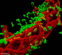
That may change with the introduction of Imaris 4D modelling software from Bitplane, a company in the Andor group. This is enabling in vivo analysis of the dynamics of leukocyte migration with a high degree of spatial and temporal resolution, leading to a new understanding of TEM regulation highlighting the role of junctional adhesion molecule C (JAM-C).
Dr Abigail Woodfin and colleagues at the Centre for Microvascular Research have reported their work in this area in a paper. The centre is well known for its application of specialised imaging, including confocal intravital microscopy which allows in vivo observation of events within the microcirculation, in 3D, in real time.
“We investigated several 3D modelling platforms before we chose Imaris. As well as a comprehensive feature set and user-friendly interface, it proved the most stable when analysing very large files – which was essential”, says Dr Woodfin. “Because of this, Imaris enabled us to track the movement of leukocytes relative to the endothelial cells lining the vessel wall over many sequential time points.
“During long analyses there is always some movement in living tissue, and we used the Imaris drift correction function to correct for this. The crucial thing was that the software allowed us to convert our data into a virtual 3D object that could be fully manipulated in rotation, zoom, and intensity. This is not possible using 2D projections of Z-stacks, which is how confocal microscopy images are traditionally presented”.

