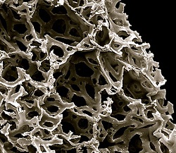
A NEW white paper from Reinnervate provides helpful information on making the switch to 3D cell culture using the company’s Alvetex scaffold. The document covers a number of different approaches to the visualisation and growth of cells in 3D, as well as the necessary daily health checks on culture growth using the technique.
Conventional 2D culture, referred to as ‘monolayer’ culture, is relatively easy to observe by traditional microscopy. The cells are almost transparent, and conveniently arranged in a single plane.
3D culture, by comparison, presents numerous imaging challenges. These are outlined in the white paper, ‘Review of imaging techniques compatible with 3D culture of cells‘, available from Reinnervate.
Alvetex is a polystyrene scaffold with filaments (interconnects) of about 13um diameter and voids of 40um, supplied as a 200um-thick membrane. Under conventional microscopy, the lack of depth of field and optical diffraction from the scaffold structure combine to make cell imaging tricky.
Among the techniques available for 3D cell imaging, discussed in the white paper, are live cell imaging, fluorescent marker analysis, confocal analysis, and electron microscopy.

