BEFORE using nanoparticle tracking analysis with the Nanosight LM-10, oncology researchers at the Department of Pediatrics at Weill Cornell University, USA, measured exosome size by the only other available method – electron microscopy.

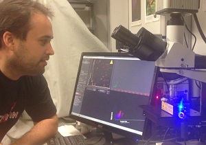
Dr Peinado Selgas using the NanoSight LM-10 at Weill Cornell Medical College, New York
In research recently published in Nature Medicine, Dr Hector Peinado describes how he and senior author Dr David Lyden’s research group were able to gain better understanding and characterisation of exosomes, secreted nanoparticles from tumour cells, using Nanosight’s nanoparticle tracking analysis (NTA) technology.
“We are analysing the role of tumour-secreted exosomes in metastasis, and study how exosomes secreted from melanoma tumour cells are educating bone marrow derived progenitor cells toward a pro-metastatic phenotype.
“We also analyse the use of exosomes as biomarkers of specific tumour types and their use as prognostic factors, technology on which Cornell University has pending patents.
“We have found that the protein content per exosome is increased in metastatic melanoma patients. In addition, we have observed that metastatic cell lines also have increased protein content per exosome.
“Therefore, knowing the number of exosomes was a definitive and necessary step in our research. Before this work, we were only following qualitative changes in exosomes. Now we are able to make quantitative analyses using nanoparticle tracking analysis.
“This technology allows us to analyse millions of particles, particle by particle, in minutes giving not only numbers but also population distribution. Although the measurement of the size of the particles is not as accurate as the electron microscopy, NTA does allow us to process a large number of samples in a short time period.”
Melanoma exosomes educate bone marrow progenitor cells toward a pro-metastatic phenotype through MET – Nature Medicine18, 883-891 (2012) doi:10.1038/nm.2753
