
A NEW technique to combine two distinct microscope technologies can create sharper images of rapid processes inside a cell, say its inventors.
Scientists at the US National Institute of Biomedical Imaging and Bioengineering (NIBIB) developed the technique to apply High Resolution Optical Imaging (HROI) to improve Total Internal Reflection Fluorescence (TIRF) microscopy.
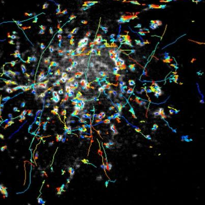
The rapid movements of Rab11 particles can be clearly imaged with the new instant TIRF-SIM microscope. Image courtesy Hari Shroff, US National Institute of Biomedical Imaging and Bioengineering
TIRF microscopy illuminates samples at an acute angle, so that light reflects back and only a thin section of the sample close to the coverslip is illuminated.
This eliminates much out-of-focus background light and creates very high contrast images.
However, conventional TIRF produces blurry images of small features within cells.
So-called super-resolution microscopy techniques have been able to improve TIRF resolution, but slow down the process so much that it has been impossible to generate sharp images of fast-moving objects.
Hari Shroff, chief of NIBIB’s High Resolution Optical Imaging (HROI) laboratory, developed a technique called instant structured illumination microscopy (iSIM) in 2013.
This can capture video at 100 frames per second, about four times as fast as most movies or online videos, but these do not have the contrast of TIRF images.
Shroff and his team designed a simple mask to block most of the illumination from the iSIM-mimicking TIRF microscope.
As a result they found they were able to observe objects moving about ten times faster than other microscopes at similar resolution.
“TIRF microscopy has been around for more than 30 years and is so useful that it will likely be around for at least the next 30” said Shroff.
“Our method improves the spatial resolution of TIRF microscopy without compromising speed – something that no other microscope can do.
“We hope it helps clarify high-speed biology so we can better understand how biological processes work”.
Using the new microscope, Shroff and his team were able to follow rapidly moving Rab11 particles near the plasma membrane of human cells.
Attached to molecular cargo transported around the cell, these particles move so fast they are blurred when imaged by other microscopes.
As with all of the microscopes developed by the Shroff team, researchers are welcome to contact the lab to try the microscope out, or to acquire free schematics of the technology.
1 comment for “Faster microscope imaging inside living cells”