NANOPARTICLE Tracking Analysis (NTA) is being employed by the Vo-Dinh lab at Duke University’s department of biomedical engineering, to help characterise metal nanoparticle construct materials for use in biosensing, imaging, and cancer therapy, reports Nanosight.

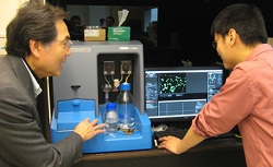
Tuan Vo-Dinh and Hsiangkuo Yuan at Duke, with their Nanosight NTA system
The main research goal of the group is to develop advanced sensors for environmental and diagnostic applications.
The research involves the design and fabricatation of metal nanoparticle constructs, ‘nanoconstructs’, such as gold nanostar platforms. Standard techniques including UV-vis spectrometry, transmission electron microscopy (TEM), Raman microscopy, and fluorometry are used to characterise these particles.
Nanoconstructs for in vivo applications, however, are in the size range from 10 to 100nm in order to be cleared from the kidney and reticuloendothelial system (RES). Size range and physiological stability are also important for optical imaging and nanodrug delivery, where it affects the dose that can be administered.
To compare the properties of nanoparticles, the researchers use NanoSight’s NTA system. This, says the company, is a major step forward from simply using TEM to look at particle shape and size, as it allows surface coating and the aggregation to also be investigated. It also provides concentration information, allowing the normalisation of particle counting comparisons.
Commenting on the benefits of using the NanoSight alongside TEM (for size) and atomic absorption spectroscopy (for mass), Professor Vo-Dinh said the ability to make characterisation particle by particle provides complementary information to the ensemble characterisation.
The group has published data on gold nanostars in the journal Nanotechnology, with another paper in Nanomedicine.
